鼻中隔
鼻中隔(Nasal septum)把人类鼻内分成两半,形成两个鼻孔。鼻中隔由筛骨,犁骨和四方形的软骨组成。
| 鼻中隔 | |
|---|---|
 Bones and cartilages of septum of nose. Right side. | |
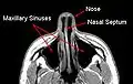 MRI image showing nasal septum. | |
| 基本 | |
| 动脉 | Anterior ethmoidal posterior ethmoidal sphenopalatine greater palatine branch of superior labial[1] |
| 神经 | Anterior ethmoidal nasopalatine nerves[1] Medial posterosuperior nasal branches of pterygopalatine ganglion |
| 淋巴 | Anterior half to submandibular nodes Posterior half to retropharyngeal and deep cervical lymph nodes |
| 标识字符 | |
| 拉丁文 | Septum nasi |
| MeSH | D009300 |
| TA98 | A06.1.02.004 |
| TA2 | 3137 |
| FMA | FMA:54375 |
| 格雷氏 | p.993 |
| 《解剖學術語》 | |
鼻中隔一般为处在鼻内中央位置,但由于天生或者后天外伤可能导致鼻中隔弯曲,堵塞空气正常经过鼻孔,一般不影响生活,但严重时可导致用鼻呼吸困难,经常流鼻血,和反复炎症,需用手术治疗。
附加圖片
 Horizontal section of nasal and orbital cavities.
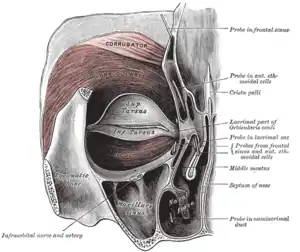 Left orbicularis oculi, seen from behind.
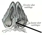 Cartilages of the nose, seen from below.
 Nasal septum
 Coronal section of nasal cavities.
 Front of nasal part of pharynx, as seen with the laryngoscope.
- MRI image showing nasal septum.
 MRI image showing deviated septum
外部連接
| 维基共享资源中相关的多媒体资源:鼻中隔 |
- Anatomy figure: 33:02-01 at Human Anatomy Online, SUNY Downstate Medical Center - "Diagram of skeleton of medial (septal) nasal wall."
- Diagram at evmsent.org
- (英文)lesson9 在韦斯利诺曼的解剖课上(乔治城大学) (nasalseptumbonescarti)
This article is issued from Wikipedia. The text is licensed under Creative Commons - Attribution - Sharealike. Additional terms may apply for the media files.